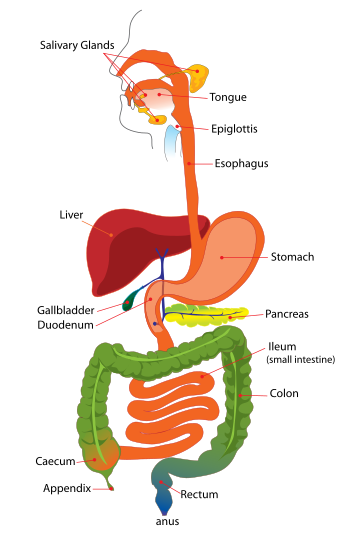
Saturday 26 May 2012

BIO 12 UNIT 3: Study Notes

IMPORTANCE OF PH IN DIGESTIVE TRACT

  • Enzymes will denature if they are not at the right pH
  • Stomach pH must be acidic to kill bacteria (HCl) and for certain enzymes to activate such as PEPSIN from pepsinogen (pH 2) [breakdown of carbohydrates]
  • Salivary Glands in the mouth must maintain a neutral pH for mechanical/chemical digestion to occur
  • Small Intestine: pH variations to match specific digestive enzyme conditions (pancreas: sodium bicarbonate) [neutralizes acidic environment]

BIO 12 UNIT 3: Study Notes

Structure and Function of Digestive System Parts


  • Mouth--> Beginning of digestive tract (where food first enters), site where chewing and initial breakdown occurs in order for food to pass through digestive tract
  • Tongue--> Causes initial food break down by moistening food to make it easier to pass through digestive tract; taste buds send nerve impulses to the brain
  • Teeth--> Chews large food particle into smaller more manageable chunks that can easily move down the digestive tract [sharp] (mechanical/physical digestion)
  • Salivary Glands--> Lubricates food, produces saliva (salivary amylase); solid with millions of secretory cells, tiny ducts that collect saliva into one centralized duct which carries saliva from gland to mouth
  • Pharynx--> Back of throat where food and air particles travel through to get to the throat (passageway)
  • Epiglottis--> Thin flap of elastic cartilage located in throat, prevents food and drink from falling down the airway (safe digestion) [prevents choking and drink from  going to the wrong bodily system]
  • Esophagus--> Located in throat close to trachea, food receptor when swallowing occurs with the aid of muscular contractions called peristalsis the food is delivered to the stomach through your esophagus; muscular tube
  • Cardiac Sphincter--> Between stomach and esophagus, prevents heartburn  (does not allow food/acid to pass back up the trachea--> pushes down to stomach)
  • Stomach--> Holding container food while it is mixed with various enzymes that further break down the food into a usable energy form, mucus protects stomach from its own acidic lining and enzymes that are responsible for chemical breakdown process, once contents are sufficiently dealt with they travel to the small intestine
  • Pyloric Sphincter--> Between small intestine and stomach, smooth muscle, allows food [chyme] to enter the small intestine (duodenum)
  • Duodenum--> The first 25 cm of small intestine where digestion of food occurs (starts at pyloric sphincter and extends towards the middle of the small intestine)
  • Liver--> Processes nutrients absorbed from small intestine, secretes bile into small intestine to aid in digestion of fat, "chemical factory"; takes in all raw materials absorbed from intestine and produces various chemicals for body functionality, breaks down and secretes drugs
  • Gall Bladder--> Stores and concentrates bile, releases to duodenum to aid with absorption and digestion of fats
  • Pancreas--> Secretes digestive enzymes into duodenum that aid in fat, carbohydrate, and protein breakdown; makes and secretes insulin (key hormone in sugar metabolism)
  • Small Intestine--> 22 foot long muscular tube, breaks down food with pancreas released enzymes and bile; peristalsis occurs here, food movement and mixture with digestive secretions from pancreas and liver; duodenum responsible for much of break down/digestion of food while the rest of the small intestine absorbs nutrients into the bloodstream; turns food semi-liquid to liquid (water, bile, mucous changes consistency --> nutrients absorb, left-over food residue liquid passes through intestine and eventually to colon)
  • Appendix--> Four inch long tube that is connected off near the small and large intestine (lower right abdomen); unknown function (scientists assume that it stores good bacteria to replenish the body after diarrheal illness or that it serves no purpose at all [evolution])
  • Large Intestine (Colon)--> 6 foot long muscular tube connecting small intestine to rectum, waste processing organ waste from digestion passes to the colon via peristalsis in liquid form, water is detracted and it is stored and released once or twice a day; stool is composed of food debris and advantageous bacteria, when colon is full of feces it empties contents into rectum to allow for elimination
  • Rectum--> 8 inch chamber connecting the colon to the anus, receives waste from the colon and stores it until it is released (evacuation); when something enters the rectum brain sensors determine whether contents should be released or not; if so, sphincters relax and contract and if not than the sphincter contracts and the feeling is eventually subsided
  • Anus--> Final element of digestive tract, 2 inch long canal composed of pelvic floor muscles and two anal sphincters, lining determines if contents are liquid, solid, or gas; sphincter muscles prevent stool from exiting at an inopportune time and pelvic floor stops stool from coming out when it should not; internal sphincter remains tight not allowing stool through (we are not aware of presence of stool) and external sphincter holds wastes inside until we go to the toilet during an urge to get rid of wastes

BIO 12 UNIT 3: Study Notes

Sequence of Events in Ovarian Cycle

1) Follicular phase: Follicle develops due to FSH released from the pituitary gland, this results in the the follicle producing estrogen and progesterone

2) Ovulation: occurs in midway of cycle (day 14), egg is released into the oviduct due to a surge in LH levels from the pituitary gland. 

3)Luteal Phase: Corpus luteum is formed from follicle remains, this produces progesterone and estrogen.If the egg is not fertilized during ovulation the corpus luteum disintegrates due to low levels of estrogen and progesterone and menstruation occurs at the end of this phase


Sequence of Events in Uterine Cycle

1) Menstrual Phase: Decreased levels of progesterone and estrogen due to break down of coprus luteum cause the endometrium to break down during days 1-5 of the cycle

2) Proliferation Phase: Endometrium rebuilds and thickens because of the developing follicle producing estrogen at the ovary. (Days 6-14)

3) Secretory Phase: Endometrium thickens and mucus is secreted, this is caused by the corpus luteum producing progesterone (Days 15-28)

http://www.youtube.com/watch?v=WGJsrGmWeKE&feature=related (video on the Menstrual Cycles)


BIO 12 UNIT 3: Study Notes

Hormone Roles In Ovarian and Uterine Cycle

1) FSH: promotes development of the follicle in the ovary
2) GNRH: causes release of FSH and LH from the pituitary gland.
3) LH: promotes development of corpus luteum in the ovary, also causes ovulation
4) Estrogen: low levels causes breakdown of endometrium, also helps to thicken uterine lining
5) Progesterone: low levels causes breakdown of endometrium, also helps to thicken uterine lining and produces mucus


BIO 12 UNIT 3: Study Notes

Positive Feedback Regulation of Oxytocin

Hormone is produced in hypothalamus and released by pituitary gland. Targets uterus and mammary glands. Increased contractions during during child birth cause more release of oxytocin which in turn causes more contractions. Increased suckling causes more oxytocin release and in turn more milk is released, more milk is available.

BIO 12 UNIT 3: Study Notes

FUNCTIONS OF ESTROGEN

  • Targets uterus and causes thickening of endometrium (PROLIFERATIVE PHASE)
  • Causes female puberty: 
    • Body Hair: pubic, armpit, legs
    • Increased body fat (curvy: hips and breasts)
    • Breast development
  • Causes eggs to mature
  • Growth of uterus and vagina


BIO 12 UNIT 3: Study Notes

Hormonal Changes as a Result of Implantation

Placenta produces HCG hormone, this helps to maintain the corpus luteum until the placenta is able to make its own progesterone and estrogen
Estrogen and Progesterone sends signals to the pituitary gland so no more follicles are matured.

BIO 12 UNIT 3: Study Notes

PATHWAY OF SPERM

Seminiferous Tubules (production site)

Epididymis (immature sperm stored here, mature)

Vas Deferens (attached to Epididymis, moves sperm away from Scrotum, stores sperm and moves it towards abdomen)

Ejaculatory Duct (paired structures that are the union of the Vas Deferens and the Seminal Vesicle sends sperm to Urethra)

None 

Urethra (Shared tube with Vas Deferens at the base of the penis; expels urine and SEMEN (sperm based fluid) 

Penis (Tip is the opening of the Urethra where the sperm exits the male body)


AN AWESOME VIDEO ILLUSTRATING THIS CONCEPT:

BIO 12 UNIT 3: Study Notes

STRUCTURE AND FUNCTION OF THE MAIN PARTS OF THE FEMALE REPRODUCTIVE SYSTEM

  • Ovaries--> Site of egg and hormone production, contains follicle and corpus luteum
  • Follicle--> Contains egg, break apart during ovulation to release egg from ovary 
  • Corpus Luteum--> Remains of the follicle after ovulation, produces hormones such as estrogen and progesterone 
  • Oviducts (fallopian tubes)--> Propels egg towards uterus; site of fertilization, contains fimbrea structures at the end that sweep eggs into tubes and towards uterus
  • Uterus--> Where implantation of fertilized egg occurs, site of embryonic development and childbirth, contains endometrium (blood vessels that become placenta or are menstruated monthly)
  • Cervix--> Entrance to uterus, holds baby in uterus, dilates during childbirth
  • Vagina--> Birth canal (where baby comes out of), exit for menstruation, site of penis reception
  • Clitoris--> Contains erectile tissue (similar to penis), important in arousal, short shaft with sensitive tip covered by fold of skin (many nerve endings)


BIO 12 UNIT 3: Study Notes

TESTOSTERONE CONTROL

---NEGATIVE FEEDBACK (Decreases production of sex hormones if the levels of sperm and testosterone are too high) [homeostasis]---

*Main control centre: HYPOTHALAMUS (brain)

*HYPOTHALAMUS releases GnHR (gonadotropin releasing hormone) targeting the ANTERIOR PITUITARY 

* ANTERIOR PITUITARY secretes:

  • LH (Lutenizing Hormone):
    • Targets the testes (Interstitial Cells that produce testosterone)
    • Increases Testosterone Production
  • FSH (Follicle Stimulating Hormone)
    • Targets testes (Seminiferous Tubules that produce sperm)
    • Increases Sperm Production

**HYPOTHALAMUS KEEPS TESTOSTERONE AND REPRODUCTIVE HORMONE LEVEL IN CHECK WITH NEGATIVE FEEDBACK WITHHOLDING PRODUCTION OF KEY HORMONES WHEN NECESSARY**


BIO 12 UNIT 3: Study Notes

FUNCTIONS OF TESTOSTERONE

1.) Aids in sperm development

2.) Puberty in males (maturation into adulthood)

3.) Results in aggressive behaviour (fighting) and aids in sexual drive [last longer in bed--> longer erection/period of sexual intercourse]

4.) Secondary Sex distinguishing characteristics (identification of males vs. females)

    • Deep voice 

    • Adams Apple

    • Muscles (gained from TESTOSTERONE)

    • Height ("growth spurt")

    • Hair growth (armpit, pubic, chest, facial, leg, arm, etc.)


BIO 12 UNIT 3: Study Notes

SPERM STRUCTURE

4 MAIN PARTS:

  • TAIL- Back of sperm, flagellum structure that propels the sperm 
  • MIDPIECE- Contains mitochondria that provide the sperm with ATP energy (functionality, motility, etc.)
  • ACROSOME- Makes up majority of the front of sperm, contains special lysosome-like digestive enzymes that will digest outer layers of the egg during fertilization
  • HEAD- The front of the sperm structure that contains the acrosome and haploid nucleus (DNA)

BIO 12 UNIT 3: Study Notes

FUNCTIONS OF SEMINAL FLUIDS

During ejaculation, the penis expels a fluid called semen made of sperm and fluids from the following three glands:

1. SEMINAL VESICLE: Secretes a high energy (fructose) fluid that serves as an energy source for sperm (motility)

2. PROSTATE: Secretes a milky, basic solution that neutralizes the acidic vagina and urethra); enhances sperm motility (sperm=more efficient and effective as a result)

3. COWPER'S: Secretes a mucus-like fluid that lubricates sperm and semen for intercourse 

BIO 12 UNIT 3: Study Notes

STRUCTURE AND FUNCTIONS OF PARTS OF THE MALE REPRODUCTIVE SYSTEM


  • Testes--> Oval shaped organs that are located in the Scrotum, make Testosterone, site for the production of sperm, contain Seminiferous tubules
  • Seminiferous Tubules--> Coiled masses of tubes that produce sperm cells (located in Testes in Scrotum)
  • Interstitial Cells--> Located in Testes in Scrotum, located between tubules, Testosterone is produced here
  • Epididymis--> Long, coiled tube that lies on the backside of each testicle, transports and stores testes produced sperm cells, site for sperm maturation
  • Vas Deferens--> Long muscular tube that travels from the Epididymis to the Urethra, transports mature sperm to the Urethra (in preparation of ejaculation) 
  • Prostate Gland--> Walnut-sized structure located below urinary Bladder and in front of rectum, contributes fluids to ejaculate (nourish sperm), urethra runs through centre of Prostate Gland 
  • Cowper's Gland--> Pea-sized structures located on the sides of the Urethra below the prostate gland, produces a clear, slippery fluid that empties into the Urethra (lubricates the Urethra and neutralizes the acidic effect of urine residue)
  • Seminal Vesicle--> Sac-like pouches attached to the Vas Deferens near the base of the Bladder, produces a fluid rich in sugar that helps sperm attain a source of energy that aid in their motility; the fluid makes up most of the volume of the male ejaculate
  • Penis--> Organ used in sexual reproduction, three parts: the root, the shaft, and the glans (head), opening of Urethra found at tip of Penis, body is cylindrical and contains three circular chambers made of special tissue, semen expelled at tip of Penis; when Penis is erect than Urethra blocks the flow of urine allowing the exclusive flow of semen to be ejaculated during an orgasm
  • Urethra--> Tube structure that carries urine from the bladder to outside the body, ejaculates semen when a male reaches an orgasm, when the Penis is erect during sexual intercourse it blocks the flow of urine and only allows semen to flow during ejaculation
  • Scrotum--> Loose, pouch-like sac of skin that hangs behind and below the Penis, contains Testicles, nerves, and blood vessels, "climate regulator" for Testes (must make sure Testes are at a slightly lower than body temperature for proper sperm maturation), special muscles in the structure's walls allow for contraction and relaxation to allow it to heat up and cool down accordingly 

BIO 12 UNIT 3: Study Notes

ROLE OF MUCUS IN GASTRIC JUICE


  • Protects stomach lining from acidic HCl (stomach would destroy itself if left in its own acidic environment)

BIO 12 UNIT 3: Study Notes

ROLE OF HCL IN GASTRIC JUICE

  • Kills bacteria, activates pepsinogen which converts to pepsin (creates appropriate pH--. 2)

BIO 12 UNIT 3: Study Notes

ROLE OF SODIUM BICARBONATE IN PANCREATIC JUICE

  • Neutralizes acidic chyme in small intestine (duodenum) [buffer], protects small intestine (changes pH to 8) 

BIO 12 UNIT 3: Study Notes

ROLE OF H2O IN DIGESTIVE JUICES

  • Acts as a solvent for the whole mixture (dissolving quality)

BIO 12 UNIT 3: Study Notes

    ENZYME                                      GLANDULAR SOURCE                          DIGESTIVE REACTION  


 Salivary Amylase 
 Salivary Glands 
 Hydrolysis of STARCH to MALTOSE (neutral pH)
 Pancreatic Amylase
 Pancreas
 Hydrolysis of STARCH to MALTOSE (basic pH)
 Lipase
 Pancreas
 Hydrolysis of LIPIDS to FATTY ACIDS and GLYCEROL (basic pH)
 Maltase
 Small Intestine
 Hydrolysis of MALTOSE to GLUCOSE (basic pH)
 Nuclease
 Pancreas
 Hydrolysis of NUCLEIC ACID to NUCLEOTIDES (basic pH)
 Peptidase
 Small Intestine
 Hydrolysis of PEPTIDES to AMINO ACIDS (basic pH)
 Trypsin
 Pancreas
 Hydrolysis of PROTEINS to PEPTIDES
 Pepsin (comes from Pepsinogen)
 Gastric Glands (stomach?)
 Hydrolysis of PROTEINS to PEPTIDES


BIO 12 UNIT 3: Study Notes


VILLUS

  • Located in small intestine, tiny finger like projections sticking out from the intestine to absorb nutrients (monomers)
  • Villi contains tiny projections called microvilli (really small and hugely increase surface area of small intestine leading to more efficient and effective absorption)
  • A villi is shaped like a tiny rod that contains a LACTEAL and CAPILLARIES 
  • CAPILLARIES absorb monosacharides and amino acids
  • LACTEALS absorb glycerides and fatty acids
  • Rod shape means villi will be surrounded by nutrients as nutrients pass by them
  • Efficient shape (rod) maximum use of space (full coverage of villi and microvilli for maximum absorption)
  • Chyme gets fully broken down into nutrients for effective absorption (back of small intestine)

BIO 12 UNIT 3: Study Notes

SMALL INTESTINE 

  • Secretes mucous (protection)
  • Organs secrete products into LUMEN of small intestine (gall bladder--> bile (fat emulsification/movement in blood stream), pancreas--> bicarbonate (buffer to counteract stomach acid), pancreas--> amylase (carbohydrate break down), pancreas--> lipase (break down of fats)) 
  • Specialized anatomical structure of each part of small intestine= absorbs different things at different parts (i.e. amino acids and fructose)
  • Active and Passive transport of nutrients (dependent on what it is, specialized structures, etc.)
  • Microvilli composition of small intestine increases surface area and rate of absorption of monomers (lacteal and capillaries)
  • Epithelial cells is where absorption occurs
Here is a brief video talking about digestion and absorption in the small intestine:


BIO 12 UNIT 3: Study Notes

SWALLOWING AND PERISTALSIS

SWALLOWING:

Movement of food from mouth through pharynx into esophagus and then the stomach

STEPS:

1.) A mass of chewed and moistened food (bolus) is pushed to back of mouth by tongue, in pharynx a swallowing reflex prevents food from entering lungs and directs it to the esophagus
2.) Muscle contractions in the esophagus (peristalsis) propel bolus
3.) Bolus passes through specific sphincter and goes into stomach (heartburn results from irritation of esophagus by leaking gastric juices from sphincter)


  • Uvula- deflects large food particles (gag reflex)
  • Soft palette- closes nasal cavity during swallowing 
  • Epiglottis- closes trachea during swallowing

PERISTALSIS: 

  • Rhythmic contractions of soft muscular esophagus lining, stomach and small intestine propels food through digestive tract

HERE IS A VIDEO OUTLINING THE STEPS IN DIGESTION:

BIO 12 UNIT 3: Study Notes

BILE AND THE EMULSIFICATION OF FATS


  • Contains salts (cholesterol) which break fats up into droplets, providing larger surface area for digestive enzymes to work on (physical/mechanical digestion)

BIO 12 UNIT 3: Study Notes


LIVER

FUNCTIONS:

1.) Detoxification of blood by removal of poisonous substances (drugs)
2.) Stores iron, fat soluble vitamins A, D, E, K
3.) Makes plasma proteins from amino acids
4.) Stores glucose as glycogen after eating and breaks down glycogen to glucose to maintain glucose concentration of blood (energy) in between meals
5.) Produce urea from hydrolysis of amino acids
6.) Coverts red blood cell hemoglobin to breakdown products which have been excreted and bile salts in bile
**7.) Produces bile (stored in gallbladder before entering small intestine where salts of bile emulsify fats)


Monday 21 May 2012

BIO 12 Week in Review...

The past week we...


  • MON- Female Reproductive System Cut + Paste, /13 assignment (completed colouring and q's, labeling, cut and paste for marks in class)
  • TUE- Notes on Estrogen, discussed marks, test, etc.
  • THU- Notes on Testosterone (finished reproduction unit), started Circulatory System with an intro. class discussion of its functions and general knowledge, Colouring sheet, structure and functions sheet of the major parts of the circulatory system (started and completed for homework), notes on the Heart, Replication of the Movement of blood in the heart/body activity (Abstract heart created with construction paper, strings)
  • FRI- Notes on Stimulation of the Heart, COOKIE DECORATING (using icing [blue and red], candy rope [blue and red], and smarties to create a heart which shows the movement of blood and its nodes--> picture can be viewed on the blog), labeling sheet of the heart and blood movement, final good-byes to Ms. Phillips (wishing her a speedy and smooth recovery, my heart goes out to you, will miss you as our BIO teacher)

Saturday 19 May 2012

BIO 12 Cookie Heart


An image of our "cookie hearts" (Above) :) 

LEGEND:

Blue side (Right atrium and Right Ventricle= deO2ated blood)
Red Side (Left Atrium and Left Ventricle= O2ated blood)
Brown Smartie (SA Node)
Yellow Smartie (AV Node)
Blue Candy (DeO2ated blood flow= Superior Vena Cava--> Right Atrium--> Pulmonary Trunk/Arteries--> Lungs)
Red Candy (O2ated blood flow= Lungs--> Left Atrium (via pulmonary arteries and veins)--> Aorta--> Body)

Tuesday 8 May 2012

Digestive System Question


  • Why do farts stink, why do we fart (causes), how do we fart?
A: 
1. Remnants of hydrogen sulfide (gaseous) and skatole contribute to the odour of farts [SULFUR] (ex. eating eggs produces stinky farts due to being rich in sulfur which is derived from bacterial production in guts). 
2. Ingesting too much air (eating/drinking too fast), insoluble food content, unfamiliar or abundant quantity of food causes more frequent farting which is the product of chemical digestive system processes: hydrolisis of carbohydartes and proteins in foods
3. Gases produced in the stomach and intestines from the aforementioned biochemical processes, gas travels up (burp) or down towards rectum and is expelled through anus (FART)